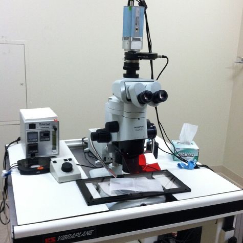
Olympus Stereo Fluorescence Imaging Microscope
By Olympus - Stereo Imaging (2025)
Function of the instrument This system is a low magnification white light and fluorescence upright imaging microscope suitable for imaging embryos, organs and tissue samples. Overview This system is a low magnification stereo microscope ideal for imaging live embryos and whole organs. It has a computer controlled motorized z-axis drive for the automated acquisition of images in multiple planes. It uses a mercury-halide fluorescence light source and filters for fluorescence imaging of nuclear stains as well as expressed fluorescent proteins. The system is equipped with a cooled monochrome CCD camera for high-sensitivity fluorescence imaging. The camera can also be used for transmitted light and oblique illumination images. Color images can be obtained using an RGB LCD electronic emission filter. A microincubation system allows for time-lapse embryo or organ culture experiments.