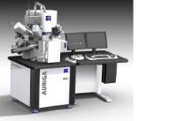
Focused Ion Beam-Scanning Electron Microscope
By Zeiss - Auriga 40 FIB-SEM (2025)
Auriga FIB-SEM - Core Research Facilities Characterization Suite (Room 154 B) - Faraday shield room. Training Requirements: 6 hours and must have demonstrated proficiency in using the JEOL 7401F FE-SEM. Ga liquid metal ion source FIB specs: 7 nm @ 30 kV, 600x - 500kx magnification, 1 pA - 20 nA for fast and precise sample modification. GEMINI® FE-SEM specs: 1.1 nm @ 20 kV, 20x - 900kx magnification, Thermal field emission type 0.1 - 30 kV, 2.5 nm @ 1 kV for Ultra high resolution. In-lens EsB® detector and STEM detector. CrossBeam® operation (milling and polishing with live SEM imaging capability). Multi-channel gas injection system for selective etching, enhanced etching, material deposition, insulator deposition. Super eucentric, fully motorized stage and dry pumping system.